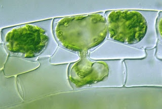
Cosmarium

Class: chlorophceae

Order: Zygnematales

Family: Desmidiaceae

Genus: Cosmarium

Cosmarium is a uni-cellular fresh water desmids occurring in ponds, rich in organic decaying matter, along with other free-floating algae. Usually it occurs in abundance in mucilaginous masses along the wall of reservoirs and water tanks especially in winter'

The cell posses a distinct median constriction called sinus. This divides them in to two distinct halves called semi- cells. Which remains join together by a connecting zone called isthmus. Each semi-cell maybe circular, elliptical or oval in shape. The cells posses a three layered transversely segmented walls with vertical poles arranged in a definite pattern. A single nucleus is found in the isthmus region. There is one or more chloroplast in each semi-cell with one or two pyrenoid.

Reproduction takes place by asexual reproduction (cell division) and isogamous sexual reproduction. Sexual reproduction results formation of zygospore.

Anabaena

Kingdom: Bacteria

Division: Cyanophyta

Class: Cyanophyceae

Order: Nostocales

Family: Nostocaceae

Genus: Anabaena

Anabaena is the filamentous form found occurring either in the single filament or as free-floating colonies or in a delicate means the stratum in permanent or semi- permanent form. Some spices of anabaena are endophytic and have within roots of circus and aquatic pteriodophyte azolla. The Trico are the same thickness through out, sometimes slightly alternated at the typical ends. They are straight cercinated or irregularly contorted concurringly with a hyaline watery sheet.

The cells are usually spherical or barrel shaped .they are typically cyanophyceae with a cell wall made up of peptidoglycogen enclosing the protoplasm. The protoplasm differentiated in outer and central colorless

The centroplasm has an incipient nuclei and the chromoplasm has phycocyrin , phcoerithrin, along with the chlorophyll on photosynthetic lamellae . The protoplasm of vegetative cells are usually filled with numerous psedovacuoles. The heterocyst’s in the filament are inter-colonial and are in a same shape as vegetative cells.

Thick walled resting cells called akinetes are found adjacent to the heterocysts.


Reproduction takes place by means of homogonium and akinetes.

Basic Structure of an Amino Acid

Basic Structure

All amino acids found in proteins have this basic structure, differing only in the structure of the R-group or the side chain.

The simplest, and smallest, amino acid found in proteins is glycine for which the R-group is hydrogen (H).


L-isomer
In proteins, only the L-isomer is found normally.

As you travel onward (from the carbonyl carbon to the amino group), the R group of L-amino acids will be on the left as shown in the molecular graphic on the right


Essential amino acids

Humans can produce 10 of the 20 amino acids. The others must be supplied in the food. Failure to obtain enough of even 1 of the 10 essential amino acids, those that we cannot make, results in degradation of the body's proteins—muscle and so forth—to obtain the one amino acid that is needed. Unlike fat and starch, the human body does not store excess amino acids for later use—the amino acids must be in the food every day.

The 10 amino acids that we can produce are alanine, asparagine, aspartic acid, cysteine, glutamic acid, glutamine, glycine, proline, serine and tyrosine. Tyrosine is produced from phenylalanine, so if the diet is deficient in phenylalanine, tyrosine will be required as well. The essential amino acids are argentine (required for the young, but not for adults), histidine, isoleucine, leucine, lysine, methionine, phenylalanine, threonine, tryptophan, and valine. These amino acids are required in the diet. Plants, of course, must be able to make all the amino acids. Humans, on the other hand, do not have all the enzymes required for the biosynthesis of all of the amino acids.

Scenedesmus

Scenedesmus is a widely distributed green algae found freely in stagnant water. It appears in pure culture in aquarium and in jar containing standing uncharged water in the laboratory.

It is colonial form with a number of cells in the coenobium (colony) being multiplies of two usually 4-8 cells. The cell are ellipsoidal or fusiform in shape and arranged in single or double series with long axis, parallel to one another so as to form a flat or curve plain. The two end cells of the coenobium may differ from the shape of the others and often teeth or spines or gelatinous bristles, which are projections of the mucilaginous cell envelope .each cell is un-nucleated and contain a single longitudinal laminate chloroplast with one pyrenoid.

Reproduction is by the formation of 2, 4,8,16 or 32 autospores by each cell in the colony. They remain laterally united to another to form a coenobium which is liberated by rupturing of the parent cell wall.


Spirulina

Spirulina is a planktonic blue green algae found growing in fresh water bodies, sewages etc. it consists of a unicellular trichrome’ which is helically or spirally twisted and hence the name Spirulina.

Each cell is typically cyanophyceae and is bounded by the cell wall enclosing the protoplasm and is differentiated in to outer pigmented chromoplasm consisting of the pigment phycocyanin and chlorophyll which give the green colour to the algae . The inner centroplasm consists of the incipient nucleus.

Reserved food materials such as cyanophytin starch, cyanophytin granules (protein) and oil globules are present in the cytoplasm

Reproduction occurs by cell division or fragmentation of the filaments.

ECONOMIC IMPORTANCE
Spirulina is excellent source of protein .vitamins and minerals, hence can be consumed as s food supplement (single cell protein) .it is also used as an animal feed due to its nutritional value.

Chlorella

Chlorella is a green algae generally found to occur as a plankton in fresh water bodies and damp walls, rocks etc. The thallus is unicellular some times growing small groups and colonies.

Each cell is small cylindrical or ellipsoidal in shape. The cell is surrounded by 3 layered cell wall. The wall shows the presence of sporopollinin. Each cell posses a single large cup shaped chloroplast which is parietal in position. The cells are unicellular. The nucleus lies in the chloroplast cup, other cell organelles like mitochondrion, ribosome etc are present.

Asexual reproduction takes place by the formation of non-motile autospores. Two to sixteen autospores are produced and they are liberated by the rupture of the parental cell wall.

Spirogyra


Spirogyra commonly called “pond silk” occurs in extensive muscles in living bright green free floating algae on the surface of stagnant ponds’ slowly running streams and fresh water streams. The thallus or plant body consists of long slender thread like un-branched filaments which are slimy to touch due to mucilage on there outer wall .
A cell enlarged
Each filament is composed of a single raw of similar cylindrical cells. Each cell consists of a cell wall made up of cellulose and protein in definite within the cell wall is a parietal layer of protoplasm enclosing a large central vacuoles. In the protoplasm is embedded 1-14 chloroplast.

Each chloroplast is in the form of a constitutes a green spiral band with toothed edges hence the name spirogyra. The chloroplast has large pyrenoids in them. There is a large single nucleus one or two nucleoli and it is suspended in the middle of the cell by cytoplasmic strands

Lateral conjugation
Reproduction occurs asexually by fragmentation of the filaments and during unfavorable conditions by the formation of walled resting spores. Sexual reproduction is by conjugation. Turning of two morphologically identical cells but physiologically dissimilar gametes.

scalariform conjugation

The Influenza (Flu) Virus

ext to the common cold, influenza or "the flu" is perhaps the most familiar respiratory infection in the world. In the United States alone, approximately 25 to 50 million people contract influenza each year. The symptoms of the flu are similar to those of the common cold, but tend to be more severe. Fever, headache, fatigue, muscle weakness and pain, sore throat, dry cough, and a runny or stuffy nose are common and may develop rapidly. Gastrointestinal symptoms associated with influenza are sometimes experienced by children, but for most adults, illnesses that manifest in diarrhea, nausea, and vomiting are not caused by the influenza virus though they are often inaccurately referred to as the "stomach flu." A number of complications, such as the onset of bronchitis and pneumonia, can also occur in association with influenza and are especially common among the elderly, young children, and anyone with a suppressed immune system.
Influenza Virus Structure

Influenza is highly contagious and is more common during the colder months of the year. Contrary to traditional belief, however, the climate itself is not directly to blame for the increase in incidence, but rather is attributable to the greater amount of time spent indoors in close proximity to other individuals during inclement weather. The influenza virus is chiefly transmitted through airborne respiratory secretions released when an infected individual coughs or sneezes. Incubation typically is from one to two days from the time of infection, and most people begin to naturally recover from symptoms within a week. The vast majority of influenza-related deaths are caused by complications of the flu rather than the actual influenza virus.

Three distinct types of influenza virus, dubbed A, B, and C, have been identified. Together these viruses, which are antigenically distinct from one another, comprise their own viral family, Orthomyxoviridae. Most cases of the flu, especially those that occur in epidemics or pandemics, are caused by the influenza A virus, which can affect a variety of animal species, but the B virus, which normally is only found in humans, is responsible for many localized outbreaks. The influenza C virus is morphologically and genetically different than the other two viruses and is generally nonsymptomatic, so is of little medical concern.

The Human Immunodeficiency Virus (HIV)

Perhaps no disease is more strongly identified with the late twentieth century than acquired immunodeficiency syndrome, commonly known as AIDS. Yet, according to a 2004 United Nations report on the state of the global AIDS epidemic, the disease has not yet begun to reduce its grip on the world population despite the fact that AIDS does not generally receive the same amount of public attention as it once did. On the contrary, infections are on the rise in many countries, including high income nations such as the United States. In 2003, nearly five million people contracted the human immunodeficiency virus (HIV) that causes AIDS, the greatest number of new infections in a single year since AIDS was first officially recognized as a disease in 1981.

Human Immunodeficiency Virus Structure

The virus responsible for HIV was first isolated in 1983 by Robert Gallo of the United States and French scientist Luc Montagnier. Since that time, a tremendous amount of research focusing upon the causative agent of AIDS has been carried out and much has been learned about the structure of the virus and its typical course of action. HIV is one of a group of atypical viruses called retroviruses that maintain their genetic information in the form of ribonucleic acid (RNA). Through the use of an enzyme known as reverse transcriptase, HIV and other retroviruses are capable of producing deoxyribonucleic acid (DNA) from RNA, whereas most cells carry out the opposite process, transcribing the genetic material of DNA into RNA. The activity of the enzyme enables the genetic information of HIV to become integrated permanently into the genome (chromosomes) of a host cell.

Plant Cell Structure

Plants are unique among the eukaryotes, organisms whose cells have membrane-enclosed nuclei and organelles, because they can manufacture their own food. Chlorophyll, which gives plants their green color, enables them to use sunlight to convert water and carbon dioxide into sugars and carbohydrates, chemicals the cell uses for fuel.

Anatomy of the Plant Cell

Bacteria Cell Structure

They are as unrelated to human beings as living things can be, but bacteria are essential to human life and life on planet Earth. Although they are notorious for their role in causing human diseases, from tooth decay to the Black Plague, there are beneficial species that are essential to good health.

Prokaryotic Cell Structure


Animal Cell Structure

Animal cells are typical of the eukaryotic cell, enclosed by a plasma membrane and containing a membrane-bound nucleus and organelles. Unlike the eukaryotic cells of plants and fungi, animal cells do not have a cell wall. This feature was lost in the distant past by the single-celled organisms that gave rise to the kingdom Animalia. Most cells, both animal and plant, range in size between 1 and 100 micrometers and are thus visible only with the aid of a microscope.

Anatomy of the Animal Cell

The lack of a rigid cell wall allowed animals to develop a greater diversity of cell types, tissues, and organs. Specialized cells that formed nerves and muscles—tissues impossible for plants to evolve—gave these organisms mobility. The ability to move about by the use of specialized muscle tissues is a hallmark of the animal world, though a few animals, primarily sponges, do not possess differentiated tissues. Notably, protozoans locomote, but it is only via nonmuscular means, in effect, using cilia, flagella, and pseudopodia.

The animal kingdom is unique among eukaryotic organisms because most animal tissues are bound together in an extracellular matrix by a triple helix of protein known as collagen. Plant and fungal cells are bound together in tissues or aggregations by other molecules, such as pectin. The fact that no other organisms utilize collagen in this manner is one of the indications that all animals arose from a common unicellular ancestor. Bones, shells, spicules, and other hardened structures are formed when the collagen-containing extracellular matrix between animal cells becomes calcified.

Animals are a large and incredibly diverse group of organisms. Making up about three-quarters of the species on Earth, they run the gamut from corals and jellyfish to ants, whales, elephants, and, of course, humans. Being mobile has given animals, which are capable of sensing and responding to their environment, the flexibility to adopt many different modes of feeding, defense, and reproduction. Unlike plants, however, animals are unable to manufacture their own food, and therefore, are always directly or indirectly dependent on plant life.

Most animal cells are diploid, meaning that their chromosomes exist in homologous pairs. Different chromosomal ploidies are also, however, known to occasionally occur. The proliferation of animal cells occurs in a variety of ways. In instances of sexual reproduction, the cellular process of meiosis is first necessary so that haploid daughter cells, or gametes, can be produced. Two haploid cells then fuse to form a diploid zygote, which develops into a new organism as its cells divide and multiply.

The earliest fossil evidence of animals dates from the Vendian Period (650 to 544 million years ago), with coelenterate-type creatures that left traces of their soft bodies in shallow-water sediments. The first mass extinction ended that period, but during the Cambrian Period which followed, an explosion of new forms began the evolutionary radiation that produced most of the major groups, or phyla, known today. Vertebrates (animals with backbones) are not known to have occurred until the early Ordovician Period (505 to 438 million years ago).

Fluorescence Microscopy of Cells in Culture

Cells were discovered in 1665 by British scientist Robert Hooke who first observed them in his crude (by today's standards) seventeenth century optical microscope. In fact, Hooke coined the term "cell", in a biological context, when he described the microscopic structure of cork like a tiny, bare room or monk's cell. Illustrated in Figure 2 are a pair of fibroblast deer skin cells that have been labeled with fluorescent probes and photographed in the microscope to reveal their internal structure. The nuclei are stained with a red probe, while the Golgi apparatus and microfilament actin network are stained green and blue, respectively. The microscope has been a fundamental tool in the field of cell biology and is often used to observe living cells in culture. Use the links below to obtain more detailed information about the various components that are found in animal cells.

Introduction to Cell and Virus Structure

At first glance, the petal of a flower or the skin on the back of a human hand may seem smooth and seamless, as if they were composed of a single, indistinct substance. In reality, however, many tiny individual units called cells make up these objects and almost all other components of plants and animals. The average human body contains over 75 trillion cells, but many life forms exist as single cells that perform all the functions necessary for independent existence. Most cells are far too small to be seen with the naked eye and require the use of high-power optical and electron microscopes for careful examination.

The relative scale of biological organisms as well as the useful range of several different detection devices are illustrated in Figure 1. The most basic image sensor, the eye, was the only means humans had of visually observing the world around them for thousands of years. Though excellent for viewing a wide variety of objects, the power of the eye has its limits, anything smaller than the width of a single human hair being able to pass unnoticed by the organ. Therefore, when light microscopes of sufficient magnifying capability were developed in the late 1600s, a whole new world of tiny wonders was discovered. Electron microscopes, invented in the mid-twentieth century, made it possible to detect even tinier objects than light microscopes, including smaller molecules, viruses, and DNA. The detection power of most electron microscopes used today, however, stops just short of being able to visualize such incredibly small structures as the electron orbital systems of individual atoms. Atoms are considered the smallest units of an element that have the characteristics of that element, but cells are the smallest structural units of an organism capable of functioning independently.

Yet, until the mid-seventeenth century, scientists were unaware that cells even existed. It wasn't until 1665 that biologist Robert Hooke observed through his microscope that plant tissues were divided into tiny compartments, which he termed "cellulae" or cells. It took another 175 years, however, before scientists began to understand the true importance of cells. In their studies of plant and animal cells during the early nineteenth century, German botanist Matthias Jakob Schleiden and German zoologist Theodor Schwann recognized the fundamental similarities between the two cell types. In 1839, they proposed that all living things are made up of cells, the theory that gave rise to modern biology.

Since that time, biologists have learned a great deal about the cell and its parts; what it is made of, how it functions, how it grows, and how it reproduces. The lingering question that is still being actively investigated is how cells evolved, i.e., how living cells originated from nonliving chemicals.

Numerous scientific disciplines—physics, geology, chemistry, and evolutionary biology—are being used to explore the question of cellular evolution. One theory speculates that substances vented into the air by volcanic eruptions were bombarded by lightning and ultraviolet radiation, producing larger, more stable molecules such as amino acids and nucleic acids. Rain carried these molecules to the Earth's surface where they formed a primordial soup of cellular building blocks.

A second theory proposes that cellular building blocks were formed in deep-water hydrothermal vents rather than in puddles or lakes on the Earth's surface. A third theory speculates that these key chemicals fell to earth on meteorites from outer space.

Given the basic building blocks and the right conditions, it would seem to be just a matter of time before cells begin to form. In the laboratory, lipid (fat) molecules have been observed joining together to produce spheres that are similar to a cell's plasma membrane. Over millions of years, perhaps it is inevitable that random collisions of lipid spheres with simple nucleic acids, such as RNA, would result in the first primitive cells capable of self-replication.

For all that has been learned about cells in over 300 years, hardly the least of which is the discovery of genetic inheritance and DNA, cell biology is still an exciting field of investigation. One recent addition is the study of how physical forces within the cell interact to form a stable biomechanical architecture. This is called "tensegrity" (a contraction of "tensional integrity"), a concept and word originally coined by Buckminster Fuller. The word refers to structures that are mechanically stable because stresses are distributed and balanced throughout the entire structure, not because the individual components have great strength.

In the realm of living cells, tensegrity is helping to explain how cells withstand physical stresses, how they are affected by the movements of organelles, and how a change in the cytoskeleton initiates biochemical reactions or even influences the action of genes. Some day, tensegrity may even explain the mechanical rules that caused molecules to assemble themselves into the first cells.

Virus Structure

Viruses are not plants, animals, or bacteria, but they are the quintessential parasites of the living kingdoms. Although they may seem like living organisms because of their prodigious reproductive abilities, viruses are not living organisms in the strict sense of the word.
Bacteriophage Structure

Without a host cell, viruses cannot carry out their life-sustaining functions or reproduce. They cannot synthesize proteins, because they lack ribosomes and must use the ribosomes of their host cells to translate viral messenger RNA into viral proteins. Viruses cannot generate or store energy in the form of adenosine triphosphate (ATP), but have to derive their energy, and all other metabolic functions, from the host cell. They also parasitize the cell for basic building materials, such as amino acids, nucleotides, and lipids (fats). Although viruses have been speculated as being a form of protolife, their inability to survive without living organisms makes it highly unlikely that they preceded cellular life during the Earth's early evolution. Some scientists speculate that viruses started as rogue segments of genetic code that adapted to a parasitic existence.

DISEASES

Communicable diseases are those caused by viruses, bacteria, and other microbes. Among the most well-known communicable diseases, otherwise known as infectious diseases, include sexually transmitted diseases (STD’s), severe acute respiratory syndrome (SARS), and leprosy.

These diseases are readily transferable, often through contact with someone carrying the disease.

They can be spread by direct human contact, through air or water, and by insects. Many health officials predict an increase in communicable diseases in the coming years, including the reemergence of diseases such as tuberculosis that were thought to have been virtually eradicated.

One cause for concern is antibiotic-resistant microbes, which make illnesses more life-threatening because existing treatments are less effective.

There is also a growing concern about the use of disease-causing microbes as weapons in the form of biological warfare or bioterrorism.

The reports of anthrax sent by mail are a well-known recent example. Smallpox is one other potential threat, but the risk of bioterrorism occurring is unknown.

HEALTH & WELLNESS

The World Health Organization defines health as a state of complete physical, mental, and social well-being and not merely the absence of disease or infirmity. Eating right, exercising, and sleeping well are all very important in the prevention of infections and diseases, but equally important is a good sense of self, a loving support network, and the potential for continued personal growth.

Many of us are not in control of the factors that cause us to become ill – whether they be genetic, environmental, or something else entirely. And too many of us do not really know how to take care of ourselves; instead we rely on doctors to “cure us.” There are many avenues we can take to improving our health; natural therapies, and both traditional and modern medicines.

But the playing field is not level, and most people cannot access or afford proper health care. Knowledge of healthy sanitation and hygiene practices and preventable diseases should be fundamental, but health education is not a given [awkwardly put] . Many people are disenfranchised because of poverty, geographic location, disability, or social stigma against certain behaviors, illnesses and diseases. Sexual health is a contentious issue around the world and ignorance about certain diseases (particularly sexually transmitted diseases such as HIV/AIDS), can cause unsafe behaviors, isolation, and premature death. The most pressing health issue today, many believe, is the education, prevention and treatment of HIV/AIDS

 
Design by Free WordPress Themes | Bloggerized by Lasantha - Premium Blogger Themes | cna certification